受賞者からのコメント
 |
ベストポスター賞を受賞して武田薬品工業株式会社 リサーチ 薬物動態研究所
|
この度は,『Proposal of a novel quantitative PCR methodology that can confirm “true cellular kinetics” in chimeric antigen receptor T cell therapy』という演題で,ベストポスター賞を賜りました.このような大変栄誉ある賞を頂き光栄に存じます.選考委員の先生方,ならびに日本薬物動態学会関係者各位に厚く御礼申し上げます.
CD19を標的としたキメラ受容体発現T細胞(chimeric antigen receptor T cell, CAR-T細胞)が,血液がんにおいて高い治療効果を示したことで,様々な抗原に対するCAR-T細胞製品の研究開発が盛んに実施されています.これまでに,quantitative polymerase chain reaction (qPCR)によって,CAR-T細胞投与数日後の一過性の血液中標的遺伝子コピー数の減衰やその後のin vivo増殖といったユニークな血液中細胞動態プロファイルが明らかとなっており[1],細胞動態と治療効果との相関性について検討が盛んになされています.細胞動態試験における血液中標的遺伝子濃度の単位としてゲノムDNA(gDNA)量に対する標的遺伝子コピー数の割合(copies/μg gDNA)が使用されていますが,CAR-T細胞投与前には化学療法による白血球除去を行うことが多く,血液中のgDNA量が,患者内および患者間で変動することが想定されます.そこで,低分子薬やバイオロジクスの薬物動態試験において一般的に使用されている単位生体内試料量における標的遺伝子コピー数(copies/μL blood)を単位としたqPCR法の構築を試みました.
一般的に,qPCRにおけるDNA抽出過程ではサンプルごとに抽出率が異なることが知られていますが,従来のqPCR法では,gDNA量に対する標的遺伝子コピー数の割合を求めれば良いため,qPCR測定プレートの各ウェルへgDNA量が均一となるようにサンプルを添加することで,抽出率のバラツキを無視することができます.また,従来法は一定量のgDNAを添加した抽出液をマトリクスとして調製した標準溶液を検量線サンプルとするため,元の生体試料中にあった標的遺伝子コピー数を求めることはできませんが,copies/μg gDNAを求めるには,PCR測定プレートの各ウェルに添加した抽出液中のコピー数を測定すれば良いので問題ありません.一方,copies/μL bloodを単位としたqPCR法では,抽出率を加味する必要があります.そこで,我々はイヌgDNAを外部標準遺伝子として各サンプルに添加することで抽出率のバラツキ補正を行いました.さらに,元の生体試料中にあった標的遺伝子コピー数を求めるために,血液に標的遺伝子をスパイクインしたサンプルを検量線試料として用いました.その結果,標的遺伝子コピー数をcopies/μL bloodの単位で示すことができるqPCR法を構築でき,その高い定量性も証明することができました.
次に,本qPCR法の有用性を検証するために,ヒト血液および白血球を除去したヒト血液にCAR-T細胞を添加し,copies/μg gDNAとcopies/μL bloodの単位でCAR遺伝子コピー数を比較しました.その結果,copies/μL bloodの単位ではいずれの試料においても添加細胞数に応じた標的遺伝子コピー数の増加が確認でき,コピー数の絶対値も生体試料間で相違ない値でした.しかし,copies/μg gDNAの単位ではヒト血液においては添加細胞数に応じた標的遺伝子コピー数の増加が確認できましたが,白血球を除去したヒト血液においては,添加細胞数に応じた標的遺伝子コピー数の増加は明確には認められませんでした.これは,CAR-T細胞を添加した際,標的遺伝子コピー数の増加に伴ってCAR-T細胞由来のヒトゲノムDNAが増加するため,ヒトgDNAの含有量が顕著に低い白血球を除去したヒト血液では,添加細胞数に応じた標的遺伝子のコピー数の増加が認められなかったからだと考えられました.このように,copies/μg gDNAを単位として用いた場合,検体に含まれるgDNA量によってCAR-T細胞の生体内での増減に対する標的遺伝子コピー数の変動を正しく評価できないため,CAR-T細胞の生体内での増減に対する標的遺伝子コピー数の変動を検体の種類によらず定量的に評価するには,copies/μL bloodを単位とした方が望ましいと考えられます.
最後に,本研究のインパクトについて述べたいと思います.冒頭に述べたユニークな細胞動態は真の動態プロファイルを表しているのでしょうか?化学療法によってgDNA量が著しく低下した状態でCAR-T細胞を投与した際,投与後初期のcopies/μg gDNAは分母が低くなるため,相対的に高くなってしまうことが考えられます.その後,血液中の白血球数が回復することでgDNA含量も元に戻るため,一過性のcopies/μg gDNAの低下が想定されます.さらに,CAR-T細胞がin vivoで増殖し始めるとcopies/μg gDNAも上がりますが,CAR-T細胞由来のgDNAが血液中gDNA量に影響するほど増殖するとCmaxを過小評価したり,投与量依存性が認められなかったりする可能性もあります.実際にそのような臨床試験結果も報告されております[2].本研究成果によって,真のCAR-T細胞動態が明らかとなり,早期に患者様の薬効を予測できる動態パラメータを見出せるものと信じております.
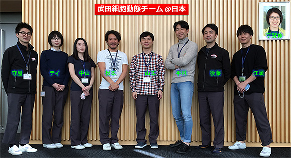
最後になりましたが,共に研究を遂行しました松本真一氏,後藤昭彦氏,宇賀神美雪氏,守屋 優氏,中山美有氏,江頭国光氏,テイネイ氏,ならびに指導を賜りました平林英樹氏,また武田薬品工業株式会社薬物動態研究所各位に,この場をお借りしまして深く感謝を申し上げます.

[1] Mueller KT et al. Blood 2017;130(21):2317-25.
[2] Raje N et al. N Engl J Med. 2019;380(18):1726-37.


